Inventory No. 35086 [1] – George III Silver Microscope
Added by Cheryl Wolfe on 28/10/2008
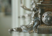
Fig. 1 Detail of the microscope on receipt.
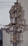
Fig. 2 Detail showing the degree of tarnishing and corrosion to iron alloy components.
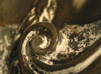
Fig. 3 Photomicrograph of cleaning product residues.
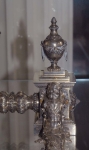
Fig. 4 Detail of cherub and urn before conservation.
A very decorative silver combined compound and simple microscope, made by George Adams, c. 1763, for King George III. The classical base and pillar support a revolving disc of eight objective lenses with each one having its own specific space. These lenses may be used as simple microscopes on one side or as part of a compound system on the other. There are two specimen stages with positioning screws, and the two mirrors serve the two methods of viewing, and it comes with a number of accessories. The microscope had previously been cleaned, but unfortunately this was not done systematically and in a very poor fashion. From the amount of corrosion and cleaning fluid residues within the detailed decorative fittings, this was probably done using a water-based silver cleaning fluid which had not been thoroughly rinsed (if at all) nor quickly dried (Figs. 1 and 2).
As a result of this the silver is tarnished very unevenly, ranging from yellow through to the violets and black, and there is a vast amount of cleaning fluid residues in the fine details of the silver (Figs. 3 and 4). The iron components are corroded, and, those components that are meant to move do not, due to build up of dirt and corrosion (in the case of iron components (Fig. 5)). There are a number of old damages to the object – small buckles and losses in the silver with some re-soldering, especially on the cherubs. There are quite a number of scratches on the silver particularly around the screws, and damage to the screw heads. Quite a number of screws are missing especially to the two figures that surmount the microscope. There is some damage to the knop of one of the cherub pillars; it is dented and has gauge marks in the silver. There are also some stress cracks in the silver.
The microscope was dis-assembled very carefully. Each section was labelled as they were removed and accompanying photographs were taken at each of these stages. Care was taken not to force any screws that would not move and the screws were kept separate, bagged and labelled to indicate which section they had come.
Each piece of the silver microscope was then surface cleaned using Hagerty Silver FoamTM in a very controlled manner using cotton wool swabs and cocktail sticks. This was repeated as necessary to obtain good level of tarnish removal. Intricate areas had to be cleaned with a combination of very fine swabs and also just the end of the cocktail stick dipped in a little of the cleaning paste. Small areas were cleaned at any one time, and, after obtaining the desired degree of tarnish removal, the residues of the foaming silver polish were removed using de-ionised water on cotton wool swabs or on soft haired brushes, and then thoroughly dried with absorbent paper towelling. Each area was then allowed to air dry further for at least 24 hours before a protective layer of microcrystalline wax was applied. Iron alloy components were cleaned of corrosion using swabs of a fine grade steel wool rolled through a little of the microcrystalline wax to act as a lubricant or with a small brass bristle brush with the wax as a lubricant. This was repeated as necessary, until a satisfactory level of corrosion removal had been achieved. Any remaining residues were removed using clean, dry cotton wool swabs.
When completed the microscope was re-assembled using the labels and photographic record as a guide (Figs. 6 – 8).
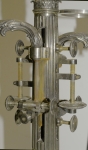
Fig. 5 Detail of corroded iron alloy components.
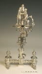
Fig. 6 Overall view after conservation.
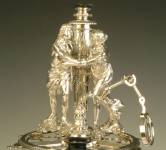
Fig. 7 Detail of figures after conservation.
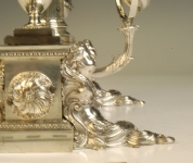
Fig. 8 Detail of cherub after conservation.
![]() For further information, see the complete conservation report
For further information, see the complete conservation report